
The gold standard of malaria diagnosis is a micrograph of a whole blood smear because the parasite spends a large part of its life in the red blood cells. For this reason, labs are working toward building point-of-care diagnostic devices which can produce a workable micrograph without the expense and hassle of a real lab-grade microscope. In first-world countries, this is a non-issue but most places where malaria is a problem are also places with a dearth of expensive laboratories for medical diagnostics.
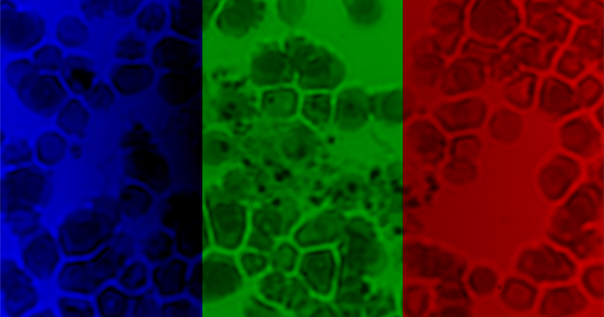
One of the issues with small devices is chromatic aberation. The different wavelengths focus at different distances from the lens creating odd color patterns. It does not change the important information content in the photo but pathologists are acclimated to viewing data a certain way. If a new device does not comform to old standards, it is almost impossible to find support in the medical field. To begin working on color correction issues, it is useful to break the image apart by color channels.
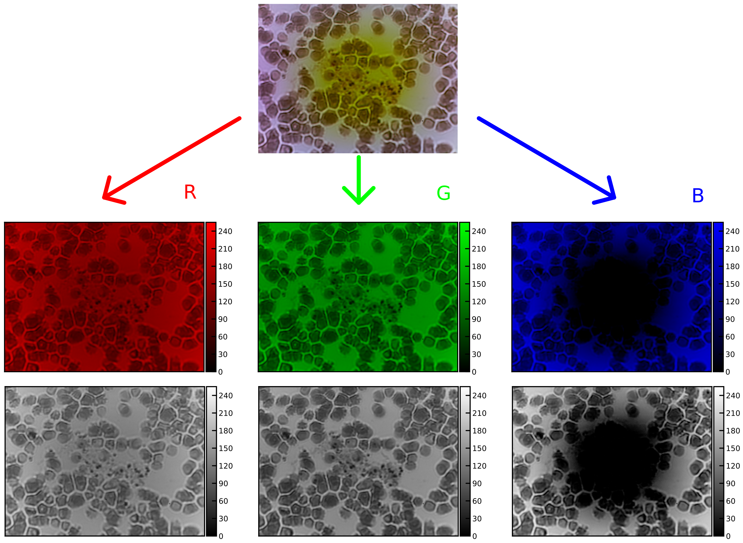
[Caption] Separation of color channels in sample image: [Top] original image unmodified, [Middle row] Red, green and blue channels separated and displayed with the remaining channels as zeros, [Bottom row] Red, green and blue channels separated and displayed in greyscale. Human eyes do not perceive different colors with equal sensitivity so displaying color channels individually in grayscale allows a person to properly perceive intensity differences.
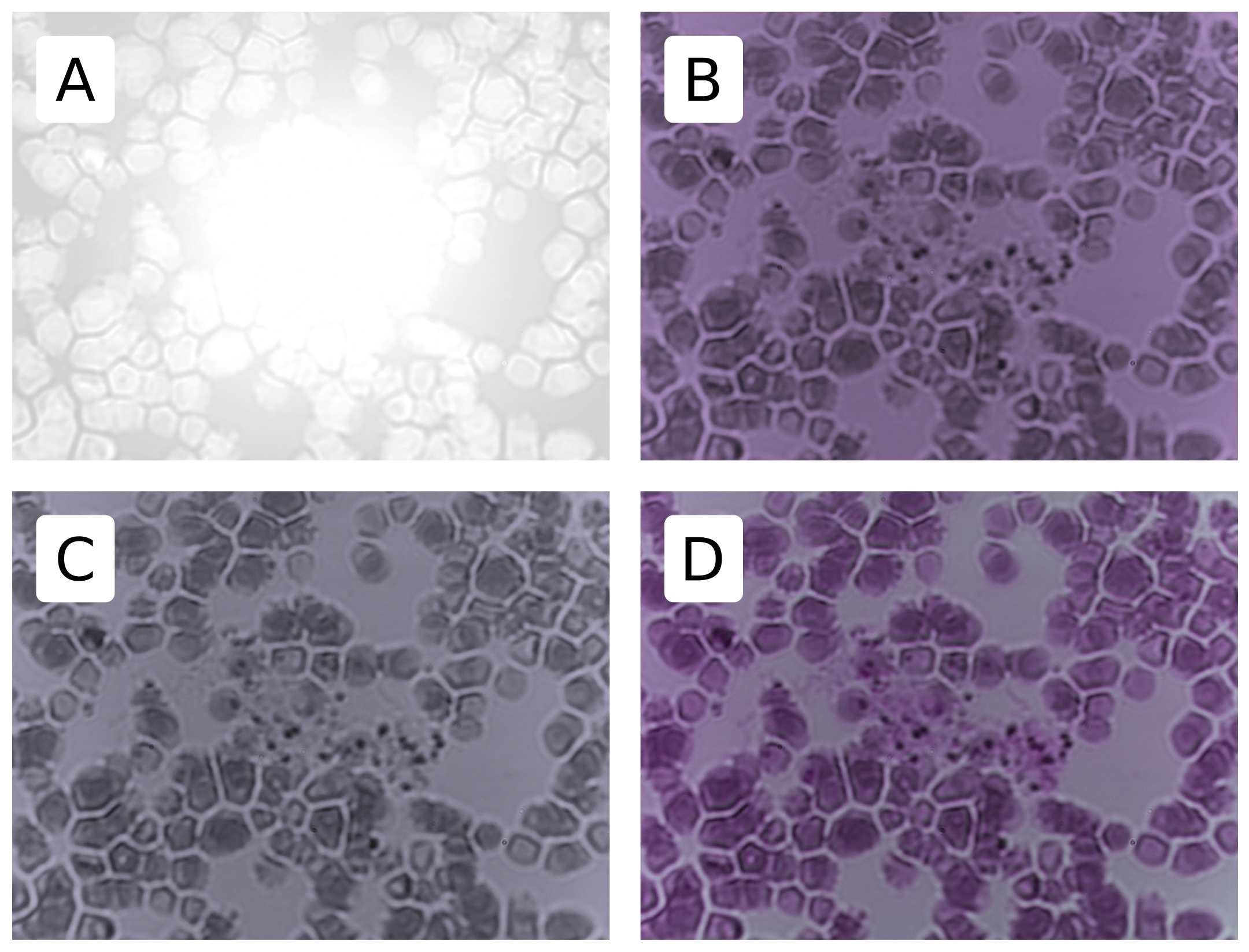
[Caption] A little creativity with color channel arithmetic and you can produce something with far fewer color disturbances.